Balo concentric sclerosis (BCS) stands as a fascinating yet perplexing entity in the realm of neurology. This rare demyelinating disorder, often considered a variant of multiple sclerosis (MS), captivates researchers and clinicians alike with its distinctive concentric ring patterns visible on brain imaging. As we delve into the intricacies of BCS, we’ll explore its unique characteristics, diagnostic challenges, and the evolving landscape of treatment options.
Table of Contents
Understanding Balo Concentric Sclerosis
First described by Hungarian pathologist József Balo in 1928, BCS is characterized by alternating layers of preserved and destroyed myelin in the brain. This pattern creates a striking “onion bulb” or “target” appearance on magnetic resonance imaging (MRI), setting it apart from other demyelinating disorders. The disease primarily affects young adults, typically manifesting between the ages of 20 and 50, though rare pediatric cases have been reported.
Clinical Presentation
The clinical presentation of BCS can vary widely, reflecting the diverse locations and extent of lesions within the central nervous system. Patients may experience a range of symptoms, including:
- Motor deficits: Weakness, spasticity, or even paralysis can significantly impact mobility and daily activities. For instance, a patient might suddenly find themselves unable to lift their arm or walk steadily.
- Sensory disturbances: Numbness, tingling, or altered sensations can affect various parts of the body, sometimes leading to balance issues or coordination problems. These symptoms might manifest as a feeling of “pins and needles” or a loss of temperature sensation in affected areas.
- Cognitive changes: Memory impairment, difficulty concentrating, or other cognitive deficits may occur, potentially affecting work or social interactions. A patient might struggle to recall recent events or find themselves easily distracted during conversations.
- Visual problems: Some patients report blurred vision or other visual disturbances, although optic neuritis is less common than in typical MS. In severe cases, this could lead to temporary vision loss or difficulties with depth perception.
- Seizures: While not as frequent, seizures can occur in some cases, adding another layer of complexity to management. These may range from focal seizures affecting a specific body part to generalized seizures involving the entire body.
Unlike classical MS, which often follows a relapsing-remitting course, BCS may present as a rapidly progressive neurological decline. This aggressive onset underscores the urgency of prompt diagnosis and intervention. However, recent studies have shown that BCS can also have a more benign course, with some patients experiencing spontaneous remission or limited progression.
Pathophysiology: Unraveling the Mystery
The hallmark of BCS lies in its unique pattern of concentric demyelination. While the exact mechanisms remain elusive, several theories have emerged to explain this phenomenon:
Cytokine-Mediated Damage
One leading hypothesis suggests that the concentric pattern results from waves of oligodendrocyte apoptosis (cell death) mediated by inflammatory cytokines. Key players in this process include tumor necrosis factor-alpha (TNF-α) and interferon-gamma (IFN-γ). These cytokines may trigger cascades of inflammation and demyelination, followed by periods of partial recovery, creating the characteristic layered appearance.
Hypoxia-Induced Tissue Injury
Another theory proposes that local tissue hypoxia plays a role in the formation of concentric lesions. This hypothesis suggests that initial inflammatory events lead to vascular compromise, creating alternating zones of tissue damage and preservation. The resulting hypoxia may trigger a protective response in some areas, leading to the preservation of myelin in concentric rings.
Diagnostic Challenges and Approaches
Diagnosing BCS presents unique challenges due to its rarity and potential overlap with other neurological conditions. A comprehensive approach combining clinical evaluation, imaging studies, and laboratory tests is essential for accurate diagnosis.
Neuroimaging: The Cornerstone of Diagnosis
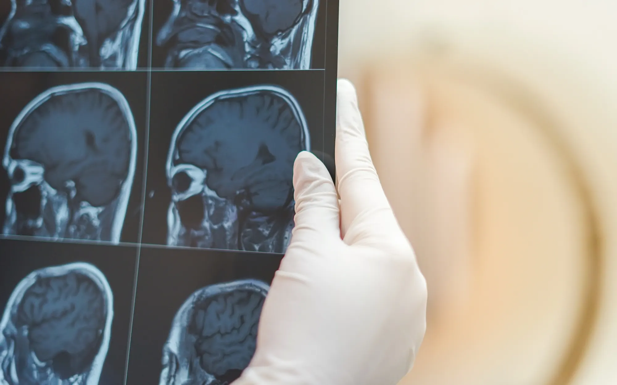
MRI stands as the gold standard for visualizing the distinctive features of BCS. T2-weighted and FLAIR sequences reveal the characteristic concentric rings, often described as “onion bulb” or “target” lesions. These images provide a visual confirmation of the disease that is crucial for diagnosis and monitoring progression.
Contrast-enhanced T1 imaging may show enhancement of active lesions, indicating ongoing inflammation. This can help differentiate between acute and chronic lesions, guiding treatment decisions.
Advanced imaging techniques, such as diffusion tensor imaging (DTI), can provide insights into the extent of axonal damage and help guide prognosis. DTI allows visualization of white matter tracts, potentially revealing areas of disruption that may not be apparent on conventional MRI sequences.
Differential Diagnosis
Distinguishing BCS from other neurological conditions is crucial for appropriate management. Key differentials include:
- Multiple sclerosis: While related, classical MS lacks the concentric ring pattern characteristic of BCS. However, some patients may present with features of both conditions, complicating diagnosis.
- Neuromyelitis optica spectrum disorder (NMOSD): Often involves optic nerves and spinal cord, with distinct antibody markers. NMOSD lesions typically lack the concentric pattern seen in BCS.
- Central nervous system lymphoma: May present with ring-enhancing lesions but lacks the alternating pattern of BCS. Careful analysis of imaging features and, if necessary, biopsy can help differentiate between these conditions.
- Acute disseminated encephalomyelitis (ADEM): Typically monophasic and more common in children, ADEM can sometimes mimic BCS in its early stages.
Treatment Approaches: Navigating Uncharted Waters
Managing BCS remains challenging due to its rarity and the lack of large-scale clinical trials. Treatment strategies often mirror those used for MS and other demyelinating diseases, focusing on immune modulation and symptomatic relief.
Acute Management
During acute exacerbations, the primary goal is to reduce inflammation and stabilize the patient’s condition. High-dose corticosteroids, such as intravenous methylprednisolone, are often the first-line treatment, aiming to suppress inflammation and hasten recovery. In severe cases or when response to steroids is inadequate, plasma exchange (plasmapheresis) may be considered to remove circulating antibodies and inflammatory mediators.
Long-term Management
For long-term disease control, clinicians often turn to disease-modifying therapies (DMTs) used in MS. These may include interferon-beta, which can help reduce relapse rates and slow disease progression, or natalizumab, a monoclonal antibody that has shown promise in preventing new lesions and relapses. Emerging therapies targeting specific immune pathways offer hope for more targeted interventions in the future.
Symptom Management and Rehabilitation
A comprehensive approach to BCS management includes addressing specific symptoms and functional impairments. Physical therapy plays a crucial role in maintaining mobility, strength, and preventing complications of immobility. Occupational therapy helps patients adapt to functional limitations and maintain independence in daily activities. Cognitive rehabilitation addresses cognitive deficits and helps patients develop compensatory strategies.
Research Frontiers and Future Directions
The rarity of BCS presents both challenges and opportunities for research. Current areas of focus include genetic studies investigating potential genetic predispositions or markers associated with BCS, advanced imaging techniques to develop more sensitive and specific imaging modalities for detecting and monitoring BCS lesions, and biomarker discovery to identify specific biomarkers in blood or cerebrospinal fluid to aid in diagnosis and prognosis.
Novel therapeutic approaches are also being explored, with researchers investigating targeted therapies that address the unique pathophysiology of BCS. These efforts hold promise for more effective and personalized treatment strategies in the future.
Conclusion
Balo concentric sclerosis remains a captivating enigma in the field of neurology. Its unique pathology, diagnostic challenges, and therapeutic hurdles underscore the importance of continued research and clinical vigilance. As we advance our understanding of BCS, we not only improve outcomes for affected individuals but also gain broader insights into the mechanisms underlying demyelinating diseases.
The journey to unravel the mysteries of BCS is far from over. With ongoing research, emerging technologies, and collaborative efforts across medical disciplines, we stand poised to make significant strides in diagnosis, treatment, and ultimately, improving the lives of those affected by this rare neurological condition.
References
- Karaarslan, E., et al. (2001). Balo’s concentric sclerosis: clinical and radiologic features of five cases. American Journal of Neuroradiology, 22(7), 1362-1367. Link
- Chen, C. J., et al. (2021). Clinical and Radiologic Features, Pathology, and Treatment of Baló Concentric Sclerosis. Neurology, 97(13), e1347-e1361. Link
- Ertuğrul, Ö., et al. (2018). Balo’s concentric sclerosis in a patient with spontaneous remission based on magnetic resonance imaging: A case report and review of literature. World Journal of Clinical Cases, 6(11), 447-454.
- Wikipedia contributors. (2024). Balo concentric sclerosis. In Wikipedia, The Free Encyclopedia. Link
- Lucchinetti, C. F., et al. (2008). The pathology of multiple sclerosis: New insights and potential clinical applications. The Lancet Neurology, 7(9), 851-861.


0 Comments