
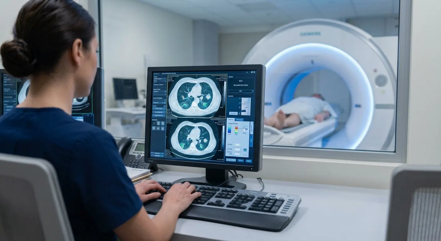
Lung cancer kills more Americans each year than breast, prostate, and colorectal cancers combined. That fact is so well-established, so thoroughly absorbed into the public health conversation, that it has calcified into a kind of shorthand: lung cancer is what happens to smokers. The connection is real — smoking accounts for roughly 80 to 90 percent of all lung cancer cases — but the shorthand has a cost. It has made lung cancer, in the minds of most people who have never smoked, someone else’s problem.
It isn’t.
Among never-smokers, lung cancer is the fifth-leading cause of cancer death worldwide, and ranks between seventh and ninth in the United States (LoPiccolo et al., Nature Reviews Clinical Oncology, 2024). The age-adjusted incidence among middle-aged never-smoking women in the U.S. runs between 15.2 and 20.8 cases per 100,000 person-years; among never-smoking men, between 11.2 and 13.7 (Wakelee et al., Journal of Clinical Oncology, 2007). Those are rates comparable to myeloma in men, or cervical cancer in women — diseases for which screening and early detection are taken seriously, funded, and recommended without controversy.
As overall smoking rates have declined and total lung cancer cases have fallen accordingly, the never-smoker share of that shrinking total has grown. Younger patients with lung cancer are disproportionately people who never smoked. And certain populations carry a burden that the aggregate statistics obscure: 57.4 percent of Asian-American women diagnosed with lung cancer, and 32.6 percent of Hispanic-American women, never smoked (Pinheiro et al., Lung Cancer, 2022). Among all lung cancer patients in seven U.S. states, never-smokers accounted for a substantial and growing fraction of cases (Siegel et al., JAMA Oncology, 2021).
None of this is reflected in current screening guidelines.
What the Guidelines Say, and What They Don’t
The U.S. Preventive Services Task Force recommends annual low-dose computed tomography screening for adults aged 50 to 80 who have smoked the equivalent of a pack a day for 20 years and who currently smoke or have quit within the past 15 years (USPSTF, JAMA, 2021). The American Cancer Society’s 2023 update follows similar logic (Wolf et al., CA Cancer J Clin, 2024). Both recommendations rest on a foundation of rigorous evidence: the National Lung Screening Trial, which enrolled 53,454 participants and demonstrated a significant reduction in lung cancer mortality with low-dose CT versus chest X-ray (NLST Research Team, NEJM, 2011), and the NELSON trial, which enrolled 15,792 participants and confirmed those findings with volume-based CT protocols (de Koning et al., NEJM, 2020).
Never-smokers are absent from both recommendations — but not because anyone ran the equivalent trials in never-smokers and found no benefit. When the USPSTF conducted its evidence review, it examined studies that did enroll never-smokers, then evaluated outcomes only in the smoker subgroups (Jonas et al., JAMA, 2021). It also excluded many studies conducted in never-smokers in East Asian countries, where the epidemiology of never-smoker lung cancer has been studied more extensively than anywhere else. The guidelines reflect a population-level mandate: identify the highest-risk group, direct resources there, and move on. That is a reasonable public health calculus. It is not the same calculation an individual never-smoker should be making about their own body.
The evidence for screening never-smokers is imperfect. There is no randomized controlled trial. What exists is a collection of observational studies, conference data, and one systematic review and meta-analysis — most of it conducted in East Asian populations where the incidence of never-smoker lung cancer is higher than in the West, and where environmental and possibly genetic drivers differ. These limitations are real and will matter to how the findings are interpreted. But the data are not nothing, and in the absence of better evidence, they are what there is.
What Screening Actually Finds
The first question worth asking is whether low-dose CT screening in never-smokers finds enough cancers to justify the exercise — and whether it finds them at a stage where treatment can actually change the outcome.
The Female Asian Nonsmoker Screening Study (FANSS), an ongoing U.S.-based lung cancer screening program enrolling Asian-American women aged 40 to 74 who have never smoked or have smoked fewer than 100 cigarettes in their lives, offers the most directly relevant American data available. In initial results from the first screening round of 963 women, 51.9 percent had benign nodules, 4.1 percent had nodules classified as likely benign but requiring follow-up, and 2.7 percent had suspicious findings. Among those with suspicious nodules, invasive lung adenocarcinoma was confirmed in 1.25 percent of all participants — 12 women — all of whom underwent surgical resection (Shum et al., Journal of Thoracic Oncology, 2025). An additional 12 women were still awaiting workup at the time of reporting.
A detection rate of 1.25 percent sounds modest until it is placed alongside the benchmarks for other screening programs that no one questions. Mammography detects breast cancer in approximately 0.4 to 1 percent of screens. Low-dose CT in the NLST — the landmark trial in smokers — detected lung cancer in roughly 1 percent of participants at the first screening round, falling to 0.34 percent in subsequent rounds among those who were initially negative (Patz et al., Lancet Oncology, 2016). In a higher-risk never-smoking population, FANSS is performing at the upper end of that range.
The meta-analysis by Triphuridet and colleagues, published in the Journal of Thoracic Oncology in 2023, pooled data from 14 studies comparing low-dose CT screening outcomes in ever-smokers and never-smokers. Thirteen of those 14 studies were conducted in East Asian countries — a significant limitation for generalizing to Western populations — but the dataset is the largest available for never-smokers. Across all studies, screening detected lung cancer in 1.00 percent (95% CI: 0.75–1.27) of ever-smokers and 0.87 percent (95% CI: 0.61–1.18) of never-smokers. In the one study from the meta-analysis that included substantial numbers of Americans — with 47 percent of never-smokers being either Asian nationals or Asian-Americans — the baseline scan detected lung cancer in 0.4 percent (95% CI: 0.3–0.6) of never-smokers. Lower than the pooled rate, but not negligible.
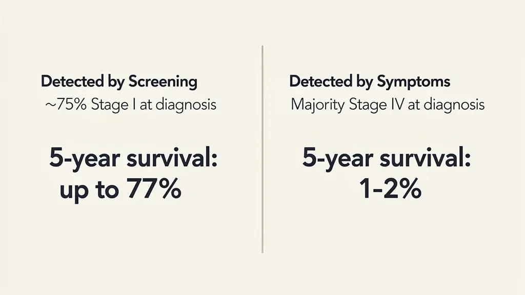
What matters as much as the detection rate is what gets detected. In the absence of screening, the majority of lung cancers in never-smokers are not caught until stage IV — the point at which five-year survival rates fall to 1 to 2 percent and the disease is, in most cases, incurable (Muallaoglu et al., Journal of BUON, 2014). In FANSS, approximately three-quarters of the cancers found on screening were at stage IA, which carries five-year survival rates as high as 77 percent. The meta-analysis found a similarly high prevalence of stage I cancers among screen-detected cases in never-smokers (Triphuridet et al., 2023). The Guangzhou Lung-Care Project, a prospective interventional study of low-dose CT screening in a mixed population of roughly three-quarters never-smokers, found consistent results in both the proportion of cancers detected and their stage distribution (Li et al., JAMA, 2025).
The difference between a stage IA diagnosis and a stage IV diagnosis is not a matter of degree. It is, in most cases, the difference between a surgical cure and a terminal prognosis.
A More Targetable Disease
There is a further reason why lung cancer in never-smokers, when caught, tends to be more tractable than lung cancer in smokers — one that has nothing to do with stage at diagnosis.
Conventional chemotherapy works by interfering with the replication of rapidly dividing cells. It does not distinguish between cancerous cells and the rapidly dividing healthy cells of bone marrow, hair follicles, and the gastrointestinal lining — which is why its side effects are what they are. Targeted therapies operate differently: they are designed to inhibit specific molecular alterations that drive a particular tumor’s growth. When a cancer carries one of those alterations and a matched drug exists, the treatment tends to be both more effective and better tolerated than conventional chemotherapy.
Never-smoker lung cancers are disproportionately driven by exactly these kinds of targetable mutations. The most important is activating mutations in the epidermal growth factor receptor gene (EGFR), which are substantially more prevalent in never-smokers than in smokers. Rearrangements in ALK and ROS1 — both of which have approved targeted therapies — are also more common in never-smoker tumors (LoPiccolo et al., 2024). The practical implication is that a never-smoker diagnosed with lung cancer is more likely than a smoker to have a treatment option specifically matched to the biology of their tumor.
The data on driver mutation prevalence comes largely from symptomatic cancers — tumors that were found because someone felt sick, not because they were screened. Most of the screening studies discussed here have not reported the mutational profiles of the cancers they detected. FANSS is an exception. All 13 lung cancers identified in the study carried actionable driver mutations: 12 had EGFR mutations, and 2 had HER2 insertions. One woman had two separate primary lung cancers, each driven by a distinct EGFR mutation (Shum et al., 2025). The sample is small, but the finding is consistent with what is known about the molecular epidemiology of never-smoker lung cancer more broadly.
The Outcome Data, Read Carefully
Early-stage detection and targetable mutations are proxies for good outcomes. The more direct question is whether never-smokers who undergo screening actually live longer — and here the evidence, while suggestive, requires careful handling.
The meta-analysis by Triphuridet and colleagues found that among people who were diagnosed with lung cancer through screening, just 3.4 percent (95% CI: 1.3–6.3) of never-smokers died from it, versus 15.0 percent (95% CI: 10.2–20.5) of ever-smokers — a difference that translated into an 87 percent lower risk of all-cause death among never-smokers with screen-detected lung cancer compared to ever-smokers in the same situation (Triphuridet et al., 2023). The Guangzhou Lung-Care Project’s long-term follow-up, presented at the European Lung Cancer Congress in 2026 and not yet published as a peer-reviewed paper, reported that people in the screening group were less than half as likely to die from lung cancer as controls over seven years of follow-up (HR 0.45, p < .001), with the difference even more pronounced in women (Li et al., ELCC 2026, conference presentation). A separate study of opportunistic low-dose CT screening in China — in which over 70 percent of participants were never-smokers — found that screened individuals were one-third less likely to die of lung cancer (HR 0.66; 95% CI, 0.54–0.80) and more than a quarter less likely to die from any cause (HR 0.72; 95% CI, 0.60–0.86) after statistical adjustment for known confounders (Wang et al., JAMA Network Open, 2023).
Those numbers are striking. They are also products of observational research, and observational research in screening studies is prone to three specific distortions that can make a test look more effective than it is.
The first is healthy user bias. People who elect to be screened are not randomly selected from the population — they are, by definition, people who sought out screening. They tend to have higher socioeconomic status, better access to care, greater health literacy, and healthier behaviors across the board. In the Chinese opportunistic screening study, 55.7 percent of the screened group carried Urban Employees Basic Medical Insurance, the better-funded insurance program for formally employed workers; only 32.1 percent of the unscreened comparison group did (Wang et al., 2023). When screened patients do better, it is not always possible to separate the effect of the screening from the effect of everything else that distinguishes people who get screened from people who don’t.
The second distortion is lead-time bias. Screening, by definition, advances the moment of diagnosis. If a cancer would have been detected in 2025 based on symptoms, and screening finds it in 2021, the patient now has four additional years of “survival” from diagnosis — even if they die at exactly the same time they would have died without screening. Five-year survival statistics, the most commonly cited metric in cancer outcomes, are particularly vulnerable to this effect: a screened patient who dies in year six looks like a survivor; an unscreened patient who dies in year two after a symptomatic diagnosis does not, even if the underlying biology was identical.
The third is length bias. Screening preferentially detects slow-growing tumors — not because it is designed to, but because fast-growing tumors are more likely to become symptomatic between screening intervals and be caught that way. If screening disproportionately finds indolent cancers that would never have caused meaningful harm, the treated patients will appear to do well — not because treatment saved them, but because their cancers were never going to kill them.
The Taiwan data illustrates this concern directly. Following increased promotion of CT screening in nonsmoking Taiwanese women, researchers observed a substantial rise in detected early-stage lung cancers — without a corresponding decline in late-stage lung cancer or lung cancer mortality (Gao et al., JAMA Internal Medicine, 2022). The pattern is consistent with overdiagnosis: the detection of real tumors that would never have become clinically significant, followed by treatment that carries its own risks and costs. Critics of low-risk screening have pointed to this data as evidence that the case for expanding screening to never-smokers is not yet made (Silvestri et al., Journal of Thoracic Oncology, 2024).
The opportunistic screening study by Wang and colleagues attempted to address these concerns more rigorously than most. Investigators matched each screened patient with a control drawn from the unscreened population on 28 baseline covariates, and used mathematical modeling to adjust for lead-time and length bias by incorporating observed lead-time estimates from clinical trials in smokers. After those adjustments, the mortality reductions held: one-third lower lung cancer mortality, more than a quarter lower all-cause mortality (Wang et al., 2023). The methodology is not perfect — residual confounding is always possible, and the lead-time adjustment borrows parameters from a different population — but it represents a more serious attempt to isolate the effect of screening than most observational studies manage.
What the evidence does not include, and what would be required to settle the question definitively, is a randomized controlled trial in never-smokers: a study in which participants are randomly assigned to receive low-dose CT screening or a control condition, and outcomes are tracked over years. The NLST and NELSON provided that standard of evidence for smokers. No equivalent trial has been conducted in never-smokers, and none appears to be on the immediate horizon.
The Risks of Screening Itself
If low-dose CT screening were free, instantaneous, and consequence-free, the absence of a randomized trial might matter less. It is none of those things, and the potential harms deserve the same scrutiny as the potential benefits.
Radiation exposure is the most direct physical risk. A single low-dose CT scan in the NLST delivered approximately 1.4 millisieverts (mSv) — roughly one-fifth the dose of a conventional diagnostic chest CT, which runs around 7 mSv (Larke et al., AJR, 2011). Modern protocols have pushed that figure lower still; current guidelines from the American College of Radiology and the American Association of Physicists in Medicine recommend an effective dose of approximately 1 mSv for a standard patient (AAPM, 2023). For context, background radiation from natural sources — cosmic rays, radon in the soil, trace radioactivity in building materials — delivers 3 to 5 mSv annually to most Americans, varying with elevation. The incremental radiation risk from a single annual low-dose CT is, by any reasonable calculation, small.
The more consequential risks are false positives and overdiagnosis. A false positive in this context means a nodule that triggers further investigation — repeat imaging, and in some cases biopsy or bronchoscopy — but turns out not to be cancer. Those follow-on procedures carry real risks: bronchoscopy and lung biopsy can cause bleeding, infection, pneumothorax, and, rarely, death (von Itzstein et al., Lung, 2019). Overdiagnosis — the detection and treatment of a genuinely cancerous lesion that would never have caused symptoms or shortened life — carries the risks of whatever treatment follows: surgery, radiation, the psychological burden of a cancer diagnosis.
The magnitude of these harms has been contested. A 2026 joint statement from the Society for Thoracic Surgeons, the American Society for Radiation Oncology, and the American College of Radiology argued that widely cited estimates of low-dose CT screening harms “contain substantial methodological flaws that contribute to the propagation of misinformation,” overstating false-positive rates, inflating the frequency of invasive follow-on procedures, and exaggerating radiation-related cancer risk (Tupper et al., Journal of the American College of Radiology, 2026). The statement reflects a genuine methodological dispute, not merely professional advocacy.
The clinical infrastructure for managing screening findings has also improved substantially since the NLST era. The Lung-RADS classification system — a standardized framework for categorizing pulmonary nodules found on low-dose CT — replaced the binary normal/abnormal read with a tiered system that assigns follow-up intervals based on nodule characteristics, reducing the proportion of findings that require immediate invasive workup. Modified Lung-RADS protocols tailored to populations with high rates of nonsmoker lung cancer have further improved the specificity of the system (Hsu et al., Academic Radiology, 2018). In a lung cancer screening program at Princess Margaret Hospital in Toronto, 84 percent of biopsied nodules were confirmed as true cancers (Wagnetz et al., AJR, 2012) — a false-positive biopsy rate far lower than critics of screening sometimes imply.
What Can Actually Be Changed
Screening is a response to a risk that already exists. The more fundamental question is whether that risk can be reduced before it requires a response.
Radon is the leading cause of lung cancer in never-smokers in the United States, responsible for up to 30 percent of never-smoker lung cancer deaths (Liu et al., Critical Reviews in Oncology/Hematology, 2024). It is a colorless, odorless radioactive gas released from subsurface rock, soil, and certain building materials, and it accumulates in enclosed spaces — basements, ground-floor rooms, poorly ventilated homes. Radon is also specifically implicated in small-cell lung cancer, which is among the most aggressive and rapidly fatal subtypes. The CDC maintains resources for home radon testing and mitigation; the intervention, where levels are elevated, is straightforward and effective.
Outdoor air pollution — particularly fine particulate matter at 2.5 microns or smaller (PM2.5) — is the second-leading cause of lung cancer cases globally and a significant contributor to never-smoker risk (Berg et al., Journal of Thoracic Oncology, 2023). Direct human-caused air pollution has declined in many parts of the developed world over recent decades, but wildfire smoke has emerged as a growing source of PM2.5 exposure in Canada, the western United States, and Australia. There is evidence that PM2.5 from wildfire smoke is more mutagenic than particles of equivalent size from ambient pollution sources (Aguilera et al., Nature Communications, 2021), and recent genomic work has begun to characterize the specific mutational signatures that air pollution leaves in the genomes of never-smoker lung cancers (Díaz-Gay et al., Nature, 2025).
Cooking fumes represent a third modifiable exposure. A 2025 meta-analysis found that never-smokers with high exposure to indoor cooking fumes were 3.68 times as likely to develop lung cancer as those with low exposure (OR 95% CI, 2.67–5.07), with the risk rising further for users of solid fuels such as wood or charcoal in enclosed spaces (OR 5.54; 95% CI, 3.15–9.72) (Hong et al., American Journal of Translational Research, 2025). Even natural gas cooking elevates indoor levels of nitrogen dioxide, particulate matter, and benzene above what the cooking itself produces (U.S. Government Accountability Office, 2025). Range hood ventilation that exhausts air outside the home — not recirculating filters — substantially reduces this exposure.
Secondhand smoke, once the dominant modifiable risk for never-smokers, has been substantially reduced by public policy in most Western countries. It remains a meaningful exposure in private spaces where smoking occurs.
The Decision That Doesn’t Make Itself
The evidence for low-dose CT screening in never-smokers is not the evidence that exists for smokers. There is no randomized trial. The observational data is concentrated in East Asian populations where never-smoker lung cancer incidence is higher than in the West, and where the environmental and genetic drivers of the disease may differ. The Taiwan data raises a legitimate concern about overdiagnosis that has not been resolved. These are not trivial limitations.
What the evidence does show, across multiple studies and populations, is that low-dose CT screening in never-smokers detects lung cancer at rates comparable to other widely endorsed screening programs; that it detects those cancers overwhelmingly at stage I, when cure rates are high; that the cancers it finds in never-smokers are more likely than smoker cancers to carry targetable driver mutations; and that the observational outcome data, even after adjustment for known sources of bias, points toward meaningful reductions in lung cancer mortality and all-cause mortality among those who are screened.
The risks of screening — radiation, false positives, overdiagnosis — are real but bounded. Radiation exposure from a modern low-dose CT is a fraction of annual background exposure. The Lung-RADS system has reduced the cascade of invasive follow-on procedures that characterized earlier screening programs. The joint statement from three major thoracic and radiology societies suggests that the harms of screening have been systematically overstated in the literature.
The gap between what the evidence supports and what the guidelines recommend is not unusual in medicine. Guidelines are instruments of population-level policy, calibrated to the highest-risk groups and the strongest available evidence. They are not designed to answer the question an individual never-smoker is actually asking: given what is known, does it make sense for me to be screened?
That question has no clean answer yet. What it has is a body of evidence that is coherent enough, and the potential consequences of an undetected early-stage lung cancer severe enough, that treating the decision as already made — by default, in the direction of not screening — is itself a choice, and not necessarily the right one.
Sources
- LoPiccolo J, Gusev A, Christiani DC, Jänne PA. Lung cancer in patients who have never smoked – an emerging disease. Nature Reviews Clinical Oncology. 2024;21(2):121–146.
- Wakelee HA, Chang ET, Gomez SL, et al. Lung cancer incidence in never smokers. Journal of Clinical Oncology. 2007;25(5):472–478.
- Pinheiro PS, Callahan KE, Medina HN, et al. Lung cancer in never smokers: Distinct population-based patterns by age, sex, and race/ethnicity. Lung Cancer. 2022;174:50–56.
- Siegel DA, Fedewa SA, Henley SJ, Pollack LA, Jemal A. Proportion of never smokers among men and women with lung cancer in 7 US states. JAMA Oncology. 2021;7(2):302–304.
- US Preventive Services Task Force, Krist AH, Davidson KW, et al. Screening for lung cancer: US Preventive Services Task Force recommendation statement. JAMA. 2021;325(10):962–970.
- Wolf AMD, Oeffinger KC, Shih TYC, et al. Screening for lung cancer: 2023 guideline update from the American Cancer Society. CA: A Cancer Journal for Clinicians. 2024;74(1):50–81.
- National Lung Screening Trial Research Team, Aberle DR, Adams AM, et al. Reduced lung-cancer mortality with low-dose computed tomographic screening. New England Journal of Medicine. 2011;365(5):395–409.
- de Koning HJ, van der Aalst CM, de Jong PA, et al. Reduced lung-cancer mortality with volume CT screening in a randomized trial. New England Journal of Medicine. 2020;382(6):503–513.
- Jonas DE, Reuland DS, Reddy SM, et al. Screening for lung cancer with low-dose computed tomography: Updated evidence report and systematic review for the US Preventive Services Task Force. JAMA. 2021;325(10):971–987.
- Shum E, Bell JL, Li W, et al. OA15.03 Female Asian Nonsmoker Screening Study (FANSS): A U.S.-based lung cancer screening program in a non-smoking population. Journal of Thoracic Oncology. 2025;20(10):S44.
- Triphuridet N, Zhang SS, Nagasaka M, et al. Low-dose computed tomography (LDCT) lung cancer screening in Asian female never-smokers is as efficacious in detecting lung cancer as in Asian male ever-smokers: A systematic review and meta-analysis. Journal of Thoracic Oncology. 2023;18(6):698–717.
- Li C, Cheng B, Li J, et al. Non-risk-based lung cancer screening with low-dose computed tomography. JAMA. 2025;333(23):2108–2110.
- Li C, Liao J, Li J, et al. ELCC 2026 – LBA5 – The impact of one-time low-dose CT screening on lung cancer mortality in a non–risk-based population: A prospective non-randomized controlled study. European Lung Cancer Congress 2026. Conference presentation; not yet peer-reviewed. https://cslide.ctimeetingtech.com/coasis_21526/attendee/confcal/presentation/list?q=Caichen+Li#presentation-abstract-194390532107
- Wang L, Qi Y, Liu A, et al. Opportunistic screening with low-dose computed tomography and lung cancer mortality in China. JAMA Network Open. 2023;6(12):e2347176.
- Patz EF Jr, Greco E, Gatsonis C, Pinsky P, Kramer BS, Aberle DR. Lung cancer incidence and mortality in National Lung Screening Trial participants who underwent low-dose CT prevalence screening: a retrospective cohort analysis. Lancet Oncology. 2016;17(5):590–599.
- Gao W, Wen CP, Wu A, Welch HG. Association of computed tomographic screening promotion with lung cancer overdiagnosis among Asian women. JAMA Internal Medicine. 2022;182(3):283–290.
- Silvestri GA, Young RP, Tanner NT, Mazzone P. Screening low-risk individuals for lung cancer: The need may be present, but the evidence of benefit is not. Journal of Thoracic Oncology. 2024;19(8):1155–1163.
- Larke FJ, Kruger RL, Cagnon CH, et al. Estimated radiation dose associated with low-dose chest CT of average-size participants in the National Lung Screening Trial. AJR American Journal of Roentgenology. 2011;197(5):1165–1169.
- AAPM Working Group on Standardization of CT. Lung Cancer Screening CT Protocols Version 6.0. American Association of Physicists in Medicine. November 9, 2023. https://www.aapm.org/pubs/CTProtocols/documents/LungCancerScreeningCT.pdf
- von Itzstein MS, Gupta A, Mara KC, Khanna S, Gerber DE. Increasing numbers and reported adverse events in patients with lung cancer undergoing inpatient lung biopsies: A population-based analysis. Lung. 2019;197(5):593–599.
- Wagnetz U, Menezes RJ, Boerner S, et al. CT screening for lung cancer: implication of lung biopsy recommendations. AJR American Journal of Roentgenology. 2012;198(2):351–358.
- Tupper HI, Shrager JB, Moghanaki D, et al. Misinformation and overestimation of computed tomography lung cancer screening harms — methodology matters: A joint statement from the Society of Thoracic Surgeons, the American Society for Radiation Oncology, and the American College of Radiology. Journal of the American College of Radiology. 2026;23(6):933–935.
- Hsu HT, Tang EK, Wu MT, et al. Modified Lung-RADS improves performance of screening LDCT in a population with high prevalence of non-smoking-related lung cancer. Academic Radiology. 2018;25(10):1240–1251.
- Liu Y, Xu Y, Xu W, He Z, Fu C, Du F. Radon and lung cancer: Current status and future prospects. Critical Reviews in Oncology/Hematology. 2024;198(104363):104363.
- Berg CD, Schiller JH, Boffetta P, et al. Air pollution and lung cancer: A review by International Association for the Study of Lung Cancer Early Detection and Screening Committee. Journal of Thoracic Oncology. 2023;18(10):1277–1289.
- Aguilera R, Corringham T, Gershunov A, Benmarhnia T. Wildfire smoke impacts respiratory health more than fine particles from other sources: observational evidence from Southern California. Nature Communications. 2021;12(1):1493.
- Díaz-Gay M, Zhang T, Hoang PH, et al. The mutagenic forces shaping the genomes of lung cancer in never smokers. Nature. 2025;644(8075):133–144.
- Hong Y, Jang KS, Xie H. Meta-analysis of the association between indoor environmental pollution and lung cancer risk in never-smokers. American Journal of Translational Research. 2025;17(8):5779–5798.
- U.S. Government Accountability Office. Gas Stoves: Risks and Safety Standards Related to Products and Ventilation. 2025. https://www.gao.gov/products/gao-25-107514
- Muallaoglu S, Karadeniz C, Mertsoylu H, et al. The clinicopathological and survival differences between never and ever smokers with non-small cell lung cancer. Journal of BUON. 2014;19(2):453–458.


