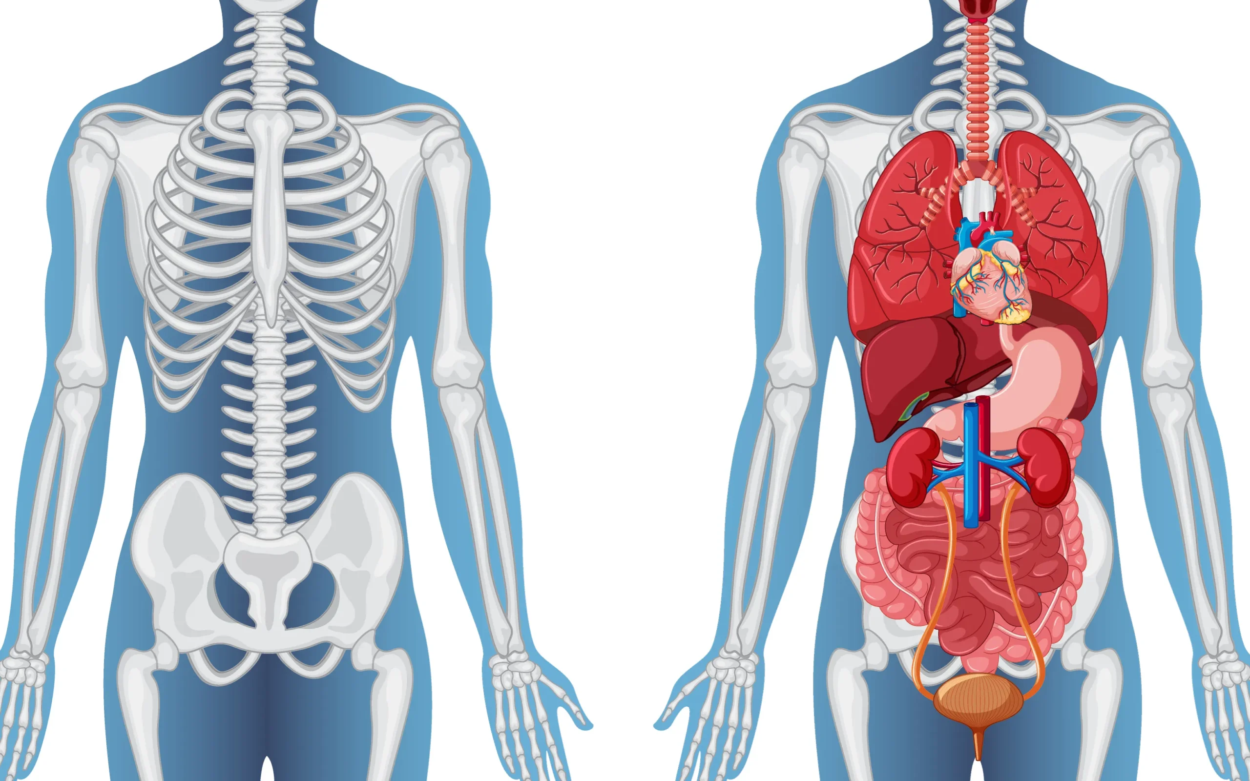
The human body is a marvel of biological engineering, with each structure finely tuned to serve specific functions. At the core of this intricate design lies the ribcage, a protective framework that safeguards our vital organs and facilitates the essential process of breathing. While most people are familiar with the standard configuration of 24 ribs arranged in symmetrical pairs, nature occasionally deviates from this blueprint. These deviations, known as anomalous rib counts, are more than mere anatomical curiosities—they can have profound implications for health, medical diagnosis, and quality of life.
Table of Contents
The Architecture of the Ribcage
To appreciate the significance of rib anomalies, we must first understand the typical structure of the human ribcage. In most individuals, the ribcage consists of 12 pairs of ribs, each with a specific role and attachment point. The first seven pairs, known as true ribs, connect directly to the sternum via costal cartilage. These ribs form a sturdy cage around the heart and lungs, providing maximum protection to these critical organs. The next three pairs, called false ribs, attach indirectly to the sternum by connecting to the costal cartilage of the rib above. This arrangement allows for greater flexibility in the lower chest, accommodating the natural expansion and contraction of the lungs during respiration. Finally, the last two pairs, aptly named floating ribs, remain unattached at their anterior ends. This design permits even more flexibility in the lower abdomen, crucial for activities like bending and twisting.
This intricate arrangement of bones and cartilage is not just a passive structure. With each breath, the ribcage expands and contracts, driven by the intercostal muscles that lie between the ribs. This rhythmic movement is essential for pulmonary function, creating the negative pressure that draws air into the lungs and the positive pressure that expels it.
Venturing Beyond the Norm: Types of Rib Anomalies
While the standard ribcage configuration serves most people well, nature occasionally experiments with variations. These anatomical deviations can manifest in several ways, each with its own set of potential implications.
Supernumerary Ribs: When More Isn’t Always Better
One of the more common rib anomalies is the presence of extra ribs, known as supernumerary ribs. These additional bones can appear in different locations along the spine, with varying frequencies and consequences.
Cervical Ribs: The Neck’s Unexpected Guest
Emerging from the seventh cervical vertebra, cervical ribs are found in approximately 0.5% to 1% of the population. These extra ribs, while often asymptomatic, can sometimes lead to a condition known as thoracic outlet syndrome. In this scenario, the additional rib can compress nearby nerves and blood vessels, resulting in pain, numbness, or weakness in the affected arm.
Lumbar Ribs: Lower Back Additions
Less common than their cervical counterparts, lumbar ribs develop in the lower back region. While they’re typically asymptomatic, these extra ribs can occasionally cause discomfort or interfere with medical procedures targeting the lower back or abdominal area.
Rib Deficiencies: When Less Poses Challenges
On the opposite end of the spectrum, some individuals are born with fewer than the standard number of ribs. This condition, known as rib absence or aplasia, is rarer than supernumerary ribs and can be associated with other developmental abnormalities.
Absent Ribs: The Missing Pieces
Missing ribs are often linked to congenital syndromes such as Poland syndrome, a rare condition characterized by the underdevelopment or absence of the chest muscle on one side of the body, often accompanied by hand abnormalities on the same side. The absence of ribs can lead to reduced protection for internal organs and may affect the mechanics of breathing.
The Origins of Rib Anomalies: A Complex Interplay
Understanding why rib anomalies occur requires delving into the intricate world of embryonic development and genetics. The formation of the human skeleton is a precisely choreographed process, guided by a complex interplay of genetic instructions and environmental factors.
Genetic Factors: The Blueprint for Development
At the heart of rib development are the homeobox (HOX) genes, a group of genes that play a crucial role in determining the body’s segmental pattern during embryonic growth. These genes act like a molecular address system, telling cells where they are in the body and what structures they should form. Mutations in HOX genes can lead to alterations in rib number or placement. For instance, specific mutations in HOX genes have been linked to the development of cervical ribs, demonstrating how small changes in our genetic code can manifest as significant anatomical variations.
Environmental Influences: Shaping Development from the Outside
While genetics provide the blueprint, environmental factors during fetal development can also influence rib formation. Maternal health conditions, such as diabetes or thyroid disorders, may affect fetal skeletal development. Exposure to certain substances, known as teratogens, during pregnancy can disrupt normal rib formation. A well-documented example is fetal alcohol syndrome, which is associated with various skeletal anomalies, including rib abnormalities. This underscores the importance of prenatal care and avoiding harmful substances during pregnancy to promote healthy fetal development.
When Anatomy Meets Medicine: Clinical Implications of Rib Anomalies
While many individuals with rib anomalies may never experience related health issues, these anatomical variations can sometimes lead to significant clinical consequences. Understanding these potential complications is crucial for both healthcare providers and patients.
Thoracic Outlet Syndrome: When Extra Ribs Cause Trouble
One of the most well-known clinical manifestations of rib anomalies is thoracic outlet syndrome (TOS), often associated with cervical ribs. TOS occurs when the nerves or blood vessels passing through the narrow space between the collarbone and the first rib become compressed. This compression can lead to a range of symptoms, including pain in the neck and shoulder, numbness or tingling in the fingers, and weakness in the hand. The presence of a cervical rib can exacerbate this compression, leading to more severe or persistent symptoms.
For individuals with TOS, everyday activities can become challenging. Simple tasks like typing on a keyboard or reaching for objects overhead may trigger pain or discomfort. In severe cases, TOS can lead to chronic pain, reduced arm function, and even vascular complications if blood flow is significantly impaired.
Diagnostic Dilemmas: When Anomalies Muddy the Waters
Rib anomalies can pose significant challenges in medical imaging and diagnosis. Supernumerary ribs, particularly when not anticipated, may be mistaken for other structures or pathologies on X-rays or CT scans. For instance, a cervical rib might be initially misinterpreted as a bone tumor or fracture, potentially leading to unnecessary worry or invasive diagnostic procedures.
Conversely, absent ribs can lead to misinterpretation of chest imaging, potentially affecting the diagnosis of conditions like pneumothorax (collapsed lung). Radiologists and clinicians must be aware of these potential variations to avoid misdiagnosis and ensure appropriate patient care.
Breathing Mechanics: When Ribs Affect Respiration
In rare cases, rib anomalies can impact respiratory function. The ribcage plays a crucial role in the mechanics of breathing, expanding and contracting with each breath to facilitate air movement in and out of the lungs. Malformed or fused ribs may reduce chest wall flexibility, potentially leading to restricted lung expansion and compromised respiratory function.
For individuals with significant rib anomalies affecting breathing, this can translate to reduced exercise tolerance, increased susceptibility to respiratory infections, or even chronic respiratory insufficiency in severe cases. Understanding these potential impacts is crucial for managing affected individuals and optimizing their respiratory health.
Navigating the Diagnostic Landscape
Accurate diagnosis of rib anomalies is essential for appropriate management and prevention of complications. Modern medical imaging techniques offer powerful tools for identifying and characterizing these anatomical variations.
X-rays: The First Line of Sight
X-rays remain the initial imaging modality for visualizing the ribcage. They provide a broad overview of rib count and structure, allowing for the identification of obvious anomalies like cervical ribs or absent ribs. However, X-rays have limitations in detailing soft tissue involvement or subtle bone abnormalities.
CT Scans: A Detailed 3D Perspective
Computed tomography (CT) scans offer a more comprehensive view of rib anomalies. By producing detailed three-dimensional images of both bone and surrounding soft tissues, CT scans can reveal intricate details of rib structure and any associated soft tissue abnormalities. This level of detail is particularly useful in planning surgical interventions or assessing the extent of thoracic outlet syndrome.
MRI: Soft Tissue Insights
Magnetic Resonance Imaging (MRI) is particularly valuable when assessing soft tissue involvement in cases of thoracic outlet syndrome. MRI can clearly visualize nerves and blood vessels, helping to pinpoint areas of compression or irritation caused by anomalous ribs.
Managing Rib Anomalies: From Conservative Care to Surgical Solutions
The approach to managing rib anomalies depends largely on the presence and severity of symptoms. For many individuals with asymptomatic rib variations, no treatment may be necessary beyond regular monitoring. However, for those experiencing complications like thoracic outlet syndrome or chronic pain, a range of treatment options is available.
Conservative Approaches: The First Line of Defense
Non-surgical interventions are typically the first line of treatment for symptomatic rib anomalies. Physical therapy plays a crucial role, focusing on improving posture, strengthening supporting muscles, and enhancing overall body mechanics. Therapists may employ techniques like stretching exercises, postural education, and ergonomic adjustments to alleviate symptoms and improve function.
Pain management is another key component of conservative care. This may involve over-the-counter pain relievers, prescription medications for more severe pain, or interventional techniques like nerve blocks. These approaches aim to reduce discomfort and improve quality of life without resorting to more invasive measures.
Lifestyle modifications can also be beneficial. Patients may be advised to avoid activities that exacerbate their symptoms, such as heavy lifting or prolonged periods with arms raised overhead. Simple changes in daily routines and work environments can often lead to significant symptom relief.
Surgical Interventions: When Conservative Measures Fall Short
In cases where conservative treatments fail to provide adequate relief, surgical intervention may be considered. The most common surgical procedure for symptomatic rib anomalies, particularly in cases of thoracic outlet syndrome, is rib resection. This involves removing the anomalous rib, typically a cervical rib, to decompress the affected nerves and blood vessels.
Rib resection is a complex procedure requiring careful planning and execution. Surgeons must navigate a delicate area filled with important nerves and blood vessels, making precision paramount. The surgery is typically performed under general anesthesia and may require a hospital stay of several days.
While rib resection can provide significant relief for many patients, it’s not without risks. Potential complications include nerve injury, bleeding, infection, and in rare cases, lung injury. As with any surgery, the decision to proceed should be made carefully, weighing the potential benefits against the risks.
Living with Rib Anomalies: Practical Advice for Patients
For individuals diagnosed with rib anomalies, knowledge and proactive management can significantly improve quality of life. Here are some practical tips for navigating life with these anatomical variations:
- Stay informed about your condition. Understanding your unique anatomy can empower you to make informed decisions about your health care and lifestyle choices.
- Maintain open communication with your healthcare provider. Regular check-ups and prompt reporting of any new or worsening symptoms are crucial for early intervention and optimal management.
- Pay attention to ergonomics and posture. This is particularly important if you have symptoms of thoracic outlet syndrome. Ensure your work and home environments are set up to minimize strain on your neck and shoulders.
- Be mindful of activities that may exacerbate symptoms. While staying active is important, you may need to modify certain exercises or activities to avoid triggering pain or discomfort.
- Explore stress-reduction techniques. Stress can exacerbate muscle tension and pain. Practices like meditation, yoga, or deep breathing exercises may help manage symptoms and improve overall well-being.
- Consider joining a support group. Connecting with others who have similar conditions can provide emotional support, practical tips, and a sense of community.
Conclusion: Embracing Anatomical Diversity
Rib anomalies, from supernumerary cervical ribs to absent floating ribs, highlight the remarkable diversity of human anatomy. While these variations can sometimes lead to clinical challenges, they also underscore the resilience and adaptability of the human body.
As our understanding of these anatomical variations grows, so does our ability to diagnose and manage them effectively. From advanced imaging techniques to refined surgical procedures, medical science continues to evolve in its approach to rib anomalies.
For individuals living with these anatomical quirks, the journey may sometimes be challenging, but it’s far from insurmountable. With the right knowledge, medical care, and lifestyle adjustments, most people with rib anomalies can lead full, active lives.
As we continue to unravel the genetic and developmental factors behind these variations, we move closer to more targeted and effective treatments. This progress not only benefits those with rib anomalies but also enriches our broader understanding of human development and anatomy.
In the end, rib anomalies remind us that there is no one-size-fits-all in human anatomy. By embracing this diversity and continuing to advance our medical knowledge, we can ensure better outcomes and improved quality of life for all individuals, regardless of their anatomical variations.
References
- Viertel, V. G., et al. (2012). Prevalence of cervical ribs in a large series of chest radiographs. American Journal of Roentgenology. https://www.ajronline.org/doi/10.2214/AJR.11.7039
- Wellik, D. M. (2007). Hox patterning of the vertebrate axial skeleton. Developmental Dynamics. https://anatomypubs.onlinelibrary.wiley.com/doi/10.1002/dvdy.21286
- Ferrante, M. A. (2012). The thoracic outlet syndromes. Muscle & Nerve. https://onlinelibrary.wiley.com/doi/10.1002/mus.23235
- Urschel, H. C., & Kourlis, H. (2007). Thoracic outlet syndrome: A 50-year experience at Baylor University Medical Center. Proceedings (Baylor University Medical Center). https://www.ncbi.nlm.nih.gov/pmc/articles/PMC1849872/


0 Comments